Visualization
Technology in Medical Education
Sherman Gorbis, D.O., F.A.A.O. 1
Richard C. Hallgren, Ph.D.2
1 Department of
Osteopathic Manipulative Medicine
2 Department of
Physical Medicine & Rehabilitation
College of Osteopathic Medicine
Michigan State University
East Lansing,
MI 48824
Abstract
Visualization technology offers the possibility of
profoundly changing the way in which osteopathic medical students assimilate
basic osteopathic principles by giving them the ability to interactively
explore biomechanical components of the musculoskeletal system, and to
investigate the effects that changes in physical properties can have upon
functionality. We are developing a
multiple volume series of computer-assisted learning (CAL) modules which use
three-dimensional, visualization technology to enhance the acquisition of
knowledge and skills necessary for clinical evaluation and treatment of the
cervical spine. These materials,
designed to serve as an adjunct to teaching strategies that faculty are currently
using, are available to students on campus through the Kobiljak Resource Center
at Michigan State University College of Osteopathic Medicine (MSUCOM) and via
the Internet (http://hal.bim.msu.edu/EdTech) to individuals and groups who are
physically removed from the MSU campus.
In addition to addressing needs in undergraduate and graduate medical
education, these osteopathic materials, delivered to users via CDROM, have been
approved for obtaining Category 1-B CME credits (http://hal.bim.msu.edu/cme).
We anticipate that the use of these materials will
facilitate understanding of static and dynamic relationships among physical
components of the musculoskeletal system, thus contributing to ongoing efforts
to develop and maintain physician, faculty, and student expertise in areas that
are uniquely osteopathic. While we have restricted our initial efforts to the
cervical and lumbar spines, future
modules will include other regions of the body. Ultimately, we will enable students to visualize the effects of
pathology as they interactively control an articulation in three-dimensional
space.
Key Words: Internet,
computer-assisted instruction, distance learning, osteopathic, CME
Acknowledgements: This study has
been supported in part by Research Grant #95-05-405 from the American
Osteopathic Association, Chicago, IL.
Address
reprint requests to:
Richard Hallgren, Ph.D.
Department of Physical Medicine & Rehabilitation
Michigan State University
East Lansing, MI
Telephone: 517-355-4674
FAX: 517-432-1339
E-Mail: hallgren@msu.edu
Web: http://hal.bim.msu.edu
Background
The Internet has facilitated the dissemination of scientific
information throughout the world. Two
of the more notable projects on the Internet are: the National Library of
Medicine’s (NLM) Visible Human Project (http://www.nlm.nih.gov/research/visible),
a collection of on-line digital images of complete male and female cadavers for
use in medical research; and the Human Genome Project’s
(http://www.er.doe.gov/production/ober/hug_top.html) map of the human genome,
that biological sum of characteristics that makes each of us individually
unique.
The Internet has the potential to profoundly change how we
teach because it provides medical educators with increasing opportunities to
deliver interactive technology to a target audience both locally and over large
distances. There are several reasons
why we think that the Internet will have a long-term impact upon medical
education: 1) The Internet is based upon a client/server model that facilitates
the creation and updating of educational materials; 2) The Internet is able to provide transparent delivery of
information to different types of computers;
3) The amount of money that is being invested in Internet infrastructure
helps ensure that it will not be a passing pedagogical fad; 4) The Internet is
capable of providing valuable health-sciences information to remote sites.
Unfortunately, the simple presentation of information is not
a sufficient condition for learning to occur.
Effective learning tools must engage the student, causing them to
assimilate new information and to construct meaning from it in terms of what
they already know. While computer-based learning modules (CBLMs) offer distinct
advantages over material in printed form, commercial software has not
specifically addressed the unique needs of an osteopathic medical student to
visualize and understand concepts that are space/time dependent. Historically, visualization technology has
been used in two separate and distinct environments. The scientific and
engineering community has used it to convey information to a viewer. The entertainment industry has used it to
engage a viewer. The perceived utility
of visualization techniques took a quantum leap forward when the entertainment
industry realized that computers could be used to create special effects in
movies. Shortly after that, the
scientific community realized that there was potential for not only presenting
information but also for holding a viewer's attention while it was being
presented. Unfortunately, until
recently, the cost of hardware and software required to develop and deliver a
sophisticated visualization application placed these tools beyond the reach of
the average educator. But now, with
increasing computational speed and decreasing cost, what we could only dream of
doing on a personal computer in the mid-1980s has now become reality. An image that once took 20 minutes to render (10) can now be visualized in a fraction of a second. This means that educators can focus on
content rather than being consumed with the mechanics of the delivery system.
Methods
Computer-based instruction is a proven method for delivering
high-impact, interactive multimedia presentations that makes the educational
process more enjoyable for students. Research has produced convincing evidence
that students who receive a simultaneous presentation of verbal and visual
information perform better on problem-solving tests than do students who only
receive a verbal explanation(3), and demonstrates increased comprehension and long-term retention of materials(4,5). Students typically
use materials such as these: a) as an advanced organizer to acquire basic
concepts; b) as a supplement to lecture materials; c) as a tool for
review. It has been found that when
materials are viewed before class, time spent in class is better used to
synthesize information rather than merely to obtain facts.
In order to provide students with a media-rich, interactive
environment we use a combination of two or more of the following: text,
graphics, images, video, audio, animation, and simulation. Animated "gif" files are used for
free-running animations; Java-based programs are used for those instances where
an interactive animation and/or simulation is desired; Quicktime video and
audio are used as a tool for reviewing selected parts of demonstrations
performed during lectures; Virtual Reality Modeling Language (VRML) is used to
interactively view anatomy and morphology of selected structures.
The instructional modules work with computers that are
readily available to most medical students.
The software is designed to execute on a 133 MHz Pentium PC computer
with 32 Mbytes of RAM memory, 16 bit color at 800X600 resolution, 100 Mbytes of
free disk space, a sound card, and a CD-ROM player. For purely pragmatic reasons, we have elected to use Internet
Explorer 4.0 for the user interface and either Windows 95 or NT 4.0 as the
preferred operating system. The user
needs only to be able to successfully navigate a windows-based interface using
a mouse. For those users off campus who
may not have access to a high-speed Internet connection, the preferred modality
of distribution is a CDROM.
Content
Palpatory criteria for identifying and evaluating somatic
dysfunction within the musculoskeletal system have been described and taught in
many different ways. While osteopathic
physicians may sometimes differ in their use of these criteria in diagnosis and
as guides to treatment, there is general agreement on the importance of
identifying pathology and restoring normal function (6). Accurate knowledge
and detailed understanding of normal human morphology and kinesiology is
essential for identifying pathology and restoring normal function. Computers, by themselves, have not been
proven to be superior to lectures and/or textbooks in transmitting purely
factual information (7). Consequently, basic morphology can probably
still be best learned from a textbook.
However, the impact that structure has upon dynamic function is
difficult to appreciate using static media.
By merging morphologic and kinematic data into a computer-generated,
three-dimensional animation model, we are able to enhance a student’s ability
to visualize the impact that pathology can have upon function within the
musculoskeletal system (8). One area that is often difficult for
students to grasp has to do with the impact of vertebral morphology upon
coupled motion mechanics. Coupled
motion is a term used to describe a predictable secondary movement that occurs
as a result of some primary movement (9,10). Turning a bolt (a primary movement) causes it to move in or
out (a secondary movement) of a threaded hole.
There are several physiological examples of coupled motion mechanics
that are important for an osteopathic physician to know and understand. One of the most important of these occurs in
the lumbar spine where axial rotation is coupled with sidebending as a
consequence of the physical orientation of the articular facets (11,12). In the absence of
dysfunction, neutral mechanics (Type I) are characterized by coupled movement
of side-bending and rotation to opposite sides. Nonneutral mechanics (Type II) are characterized by coupled
movement of sidebending to the same side as 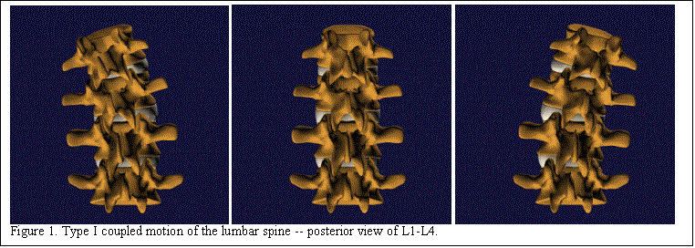
rotation. Coupled patterns of motion are complex and not easily visualized
with static pictures, but an animation sequence greatly enhances student
comprehension. Figure 1 shows three
frames representing Type I coupled motion of the lumbar spine. The left panel represents full right passive
rotation; the center panel represents the neutral position; and the right panel
represents full left passive rotation.
Notice that rotation to the left results in coupled sidebending to the
right, and that rotation to the right results in coupled sidebending to the
left.
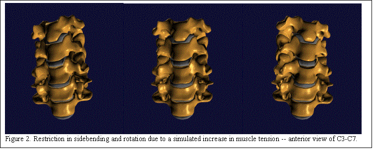
While some topics lend themselves to simple animation, other topics lend themselves to simulation.13
For example, an
asymmetric change in tension between contralateral muscles produces detectable
differences in palpable resistance to rotation when opposing directions are
compared. Physically, this results in
decreased range of motion (ROM) that an osteopathic physician can detect during
a physical examination. Figure 2 shows
an example of restricted motion that is a consequence of simulated asymmetry in
muscle tension. It is extremely
difficult to detect this restriction when looking at the static set of three
images. However, if one were to go to
our Web site
(http://hal.bim.msu.edu/edtech/cervical/biomechanics/lower/page_3.html) and
view the animated sequence, it would be clear that left sidebending is restricted. One would then gain a better appreciation
for the effectiveness of our modules to illustrate musculoskeletal dysfunction.
Quite simply, our strategy in the development of this
technological tool has been to exploit the number-crunching power of computers
to generate representations of 3-dimensional structures in areas that are
difficult to understand without the aid of animation and/or simulation, and to
use the Internet to provide access to these materials both on campus and at
distance locations.14
Conclusions
Visualization technology, one component within the
discipline of medical informatics, has the potential to facilitate student
understanding of static and dynamic relationships among physical components of
the musculoskeletal system. By properly
integrating these materials with traditional undergraduate educational
modalities such as lectures and laboratories, we hope to facilitate student
understanding of basic osteopathic principles.
With a better understanding of basic osteopathic principles, we
anticipate that our students will score higher on Osteopathic National Board
Examinations as well as feel more comfortable applying these principles in
patient care. Like most osteopathic
medical schools who rely upon community-based resources for clerkship and
graduate osteopathic medical education, MSUCOM also has a responsibility to
support its trainees (through distance learning opportunities), and trainers
(through faculty development). While
the decentralized model of osteopathic medical education provides real-world
training for students and residents, it presents challenges for administration
of the curriculum, and assuring quality and equal access to faculty and
students across the different hospitals that participate in the MSUCOM Statewide
Campus System (SCS).
We believe that by using computers as tools to implement
medical informatic applications, we will be able to provide students with the
ability to access information from a wide range of sources 24 hours per
day. This will be especially beneficial
for hospital staff who have varying work schedules, time restrictions, and
geographic constraints. Increased
implementation of computers into medical education is not the goal, but rather
a means to facilitate the educational experience of our students, thus
enhancing the quality of osteopathic medical education across the entire
educational spectrum.
References
1. Arrott M, Latta S.
Perspectives on Visualization.
IEEE Spectrum, pp. 61-65, September, 1992.
2. Hallgren RC, Reynolds HM, Soutas-Little RW, Hubbard RP,
Rechtien JJ. Three-Dimensional Analysis and Display of Sequential Position Data in
the Lumbar Spine. Journal of
Clinical Engineering, 13(1):51-57, 1988.
3. Mayer RE. Multimedia
Learning: Are We Asking the Right Questions? Educational Psychologist, 32(1), 1-19, 1997.
4. Henry JB. Computers
in Medical Education: Information and Knowledge Management, Understanding and
Learning. Human Pathology,
21(10):998-1002, 1990.
5. Jaffe CC, Lynch
PJ, Smeulder, SMW. Hypermedia Techniques for Diagnostic Imaging Instruction: Videodisk
Echocardiography Encyclopedia.
Radiology, 171:475-480, 1989.
6. Greenman PE. Principles of Manual Medicine. 2nd edition, Baltimore, Williams &
Wilkins, 1996.
7. Frisse ME. The
Case for Hypermedia. Academic
Medicine, 65:17-19, 1990.
8. Hallgren RC,
Reynolds HM. Computer Display of Multidimensional Biomedical Data. Journal of Clinical Engineering,
17(3):235-243, 1992.
9. Dowling DJ. Spinal Motion. In: An Osteopathic Approach to Diagnosis and Treatment, DiGiovanna EL
and Schiowitz S (eds). 2nd edition, New York, Lippincott-Raven, 1997.
10. Kuchera, ML. Examination and Diagnosis: An Introduction. In: Foundations
for Osteopathic Medicine, Ward, RC (executive ed). Baltimore, Williams
& Wilkins, 1997.
11. Panjabi MM,
Oxland T, Takata K, Goel V, Duranceau J, Krag, M. Articular Facets of the Human
Spine. Spine, 18(10):1298-1310,
1993.
12. Holmes A, Wang C,
Han ZH, Dang, GT. The Range and Nature of Flexion-Extension
Motion in the Cervical Spine.
Spine, 19(22):2505-2510, 1994.
13. Hallgren RC,
Gorbis S. Utilization of the Internet to Deliver Educational Materials to Health
Care Professionals. Journal of Clinical Engineering, 22(6):413-418, 1997.
14. Winn W. Learning
in Hyperspace.
http://healthlinks.washington.edu/ideal/webpaper.html, 1997.